Equipment
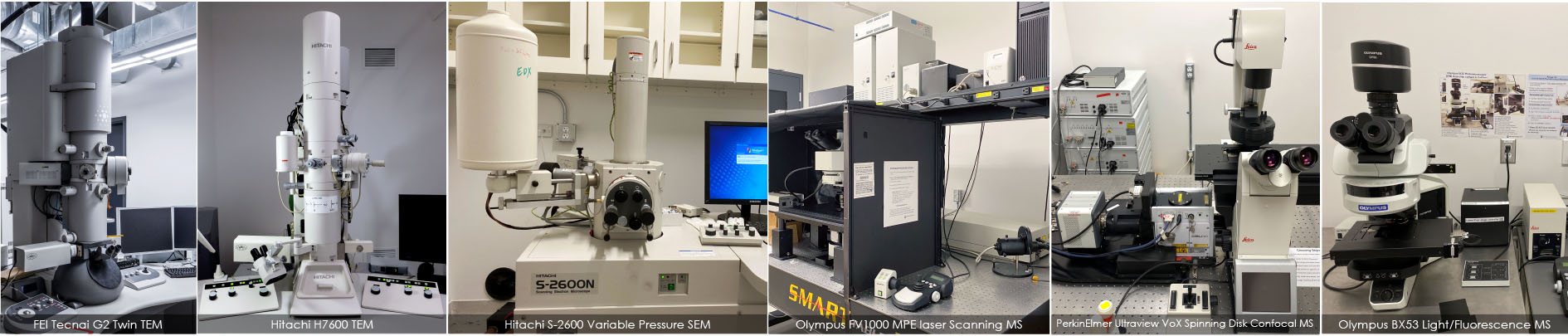
The UBC Bioimaging Facility is equipped with 4 electron microscopes (2 SEMs and 2 TEMs), 2 advanced fluorescence microscopes (confocal/multi-photon, and spinning disk), cryo-fixation (High pressure freezer, Vitrobot) and EM processing equipment (microwave, critical point dryer, coaters).
For light and electron microscopy, one vibratome and two ultramicrotomes are available for producing thick and thin sections. For image processing and analysis, we have two computer workstations with a variety of software packages installed, such as AMIRA, Dragonfly, Volocity, Inspect 3D, Photoshop, IMOD, and Fiji/ImageJ. A laminar flow cabinet and CO2 incubator are also available to keep cell and tissue cultures healthy for live-cell imaging.
| Optical Microscopes |
| Electron Microscopes |
| Sample Processing Equipment |
| Image Processing |
Techniques
The UBC Bioimaging Facility provides specialized infrastructure from optical to electron microscopy where our users can apply various types of microscopy techniques for their research. Here is a list of the techniques that are routinely used in our facility.
| TEM Techniques |
| SEM Techniques |
| EM Sample Preparation Techniques |
| Optical Microscopy Techniques |