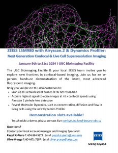
Zeiss LSM980 with Airyscan2 & Dynamics Profiler Hands-on Demo in January 2024
Zeiss is going to set up the LSM980 with Airyscan2 & Dynamics profiler for you to try ” Next Generation Confocal and Live Cell Super Resolution Imaging”. The demonstration will start on January 9th, 2024, at the UBC Bioimaging facility. You can bring your samples for the demo (live, fixed, biological and materials samples) to […]
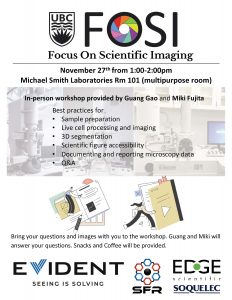
FOSI in-person workshop on November 27th 1-2pm in MSL RM 101
Focused On Scientific Imaging (FOSI) seminar is back for the month of November, and will take place on November 27th (Monday) at 1 pm in Michael Smith Laboratory (MSL) Rm 101 (multipurpose room). Guang Gao (Facility manager of LSI imaging) and Miki Fujita (Research manager at Bioimaging facility) will provide an in-person workshop about best […]
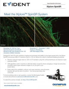
Evident/Olympus IXplore Spin SR System seminar (Nov 22) and worksho (Nov 21 to Dec 1)
Evident/Olympus will give a seminar and workshop about their IXplore Spin SR system in the week of Nov 20th. 1) Seminar about IXplore Spin SR system on November 22nd (Wednesday) 11-12pm in the Michael Smith Laboratory (MSL) Auditorium Rm102 or Zoom If you would like to join the seminar virtually, please click the link below. […]
(Untitled)
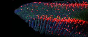
A confocal microscopy image of regenerated gill tissue in Atlantic Salmon provided by Dr. Ensiyeh Ghanizadeh-Kaserouni, a Post-doctoral Research Fellow in Brauner lab (UBC Zoology). The tissue was labelled with PCNA and NKA antibodies, and the image was acquired with the Olympus FV1000 confocal microscope. More work can be found in the recent publication.
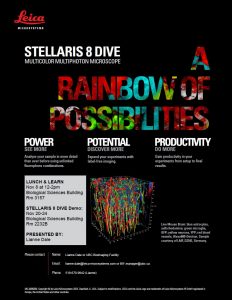
Leica Stellaris 8 Dive Multicolor Multiphoton Microscopy “Lunch and Learn” and “Demo”
We are very excited to announce that we have additional Leica events about their Stellaris 8 Dive Multicolor Multiphoton Microscope. 1) Lunch and Learn on November 8th (12-2pm) Lianne Dale from Leica will give us a talk about their demo system, Stellaris 8 Dive single photon and multiphoton combined confocal microscopy system. 2) Stellaris 8 […]
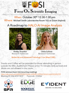
FOSI seminar on October 30th (Monday) at 12:30pm in MSL auditorium or Zoom: A Roadmap to HALO AI Image Analysis by Shelby Thornton and Olivia Sullivan
Focused On Scientific Imaging (FOSI) seminar is back for the month of October, and will take place on October 30th (Monday) at 12:30 pm in Michael Smith Laboratory (MSL) Auditorium 102 or Zoom (Hybrid). Shelby Thornton and Olivia Sullivan, will be giving a talk titled “A Roadmap to HALO AI Image Analysis”. Prior to the […]
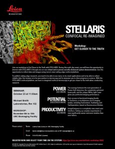
Leica Stellaris confocal microscopy seminar and workshop
We are very excited to announce that Lianne Dale from Leica will give us a talk about their Stellaris confocal microscopy system on October 23rd (Monday) from 11 am to 12 pm at the Michael Smith Laboratories (MSL) auditorium and run the workshop from November 6th to 10th at the BIF (poster is attached). During […]
(Untitled)
Dr. Belal Tafech (Hedtrich lab, Pharmaceutical Sciences) uses Perkin Elmer Spinning disk to perform live cell imaging of the restricted cilia beating of cystic fibrosis lung models built at the Hedtrich lab (left) and the Brownian motion of 10 um spherical particles in water (Right).
(Untitled)
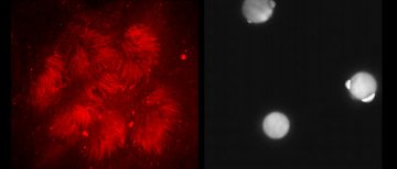
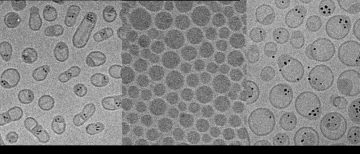
Dr. Arash Momeni (Cullis lab, Biochemistry) performs CryoTEM using the Tecnai G20 200kV TEM to examine the lipid nanoparticles. Left: 5 nm iron oxide nanoparticles and anticancer drug in lipid nanoparticles, Middle: mRNA loaded lipid nanoparticles, Right: 5 nm gold nanoparticles in lipid nanoparticles.

(Untitled)
Sean Ritter, a PhD student in Wasteneys lab (Botany), uses the PerkinElmer Spinning disk to look at cell file organization in the Arabidopsis root labelled with the plasma membrane marker LTI6b:GFP (left) and microtubule organization in the clasp-1 mutant as labelled with pUBQ1:neonGreen-TUB2 (Right).