(Untitled)
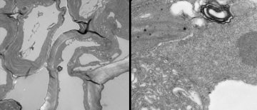
Transmission electron microscopy of Arabidopsis thaliana stem cross sections show distorted cell shapes (left image) and cytoplasmic inclusions (right image) identified in cuticle deficient mutant abcg11-3 (Bird et al., 2007. Plant J. 52 (3) 485-497). Images taken with the Tecnai Spirit 120kV TEM by undergraduate researcher Valerie Pringle in collaboration with PhD student Jessica Hancock, a […]
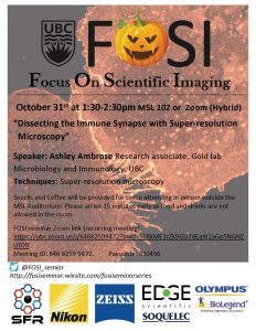
FOSI seminar on October 31st (Monday) at 1:30pm
Focused On Scientific Imaging (FOSI) seminar is back for the month of October, and will take place on October 31st (Monday) at 1:30 pm in Michael Smith Laboratory (MSL) Auditorium 102 or Zoom (Hybrid). Ashley Ambrose from Gold lab Microbiology and Immunology Department, will be giving a talk titled “Dissecting the Immune Synapse with Super-resolution Microscopy”. Prior […]
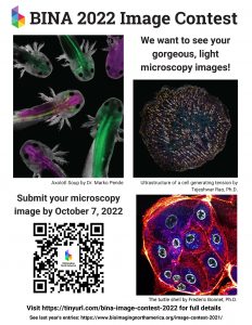
BINA 2022 Image Contest
Bioimaging North America 2022 Image Contest is open! We would like to encourage you to enter the contest. The core facility with the most submissions will win a prize. Please use a QR code below to the contest details.
(Untitled)
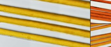
Rashika Ranasinghe, a PhD student in Dr. Darren Irwin lab Department of Zoology, studies the genomic and pigment biochemical basis of carotenoid coloration in Flameback woodpeckers in South Asia. The format of carotenoid pigment across woodpecker feathers were observed using the Olympus BX53 optilcal microscope. Left: Yellow feather of D. benghalense (Golden-backed woodpecker), Top right: […]
(Untitled)
Scanning electron micrographs of cellulose nanocrystal aerogels acquired by Lucas Andrew, a PhD student in Dr. Mark MacLachlan lab Department of Chemistry, using the Hitachi S2600 SEM.
BIF guideline July 2022
Updated on June 30th, 2022 Note: If you have any concerns, contact Miki Fujita, BIF research manager (bif.manager@ubc.ca). 1. The facility is open from 9am to 5pm Monday to Friday. * Only experienced users are permitted to use the BIF outside the business hours. Please ask BIF staff if you have any questions. A. Booking […]
(Untitled)
Please click here to register.
(Untitled)
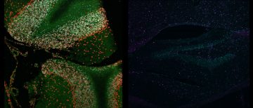
Confocal images acquired by Cristen Molzahn, a PhD student in Dr. Thibault Mayor lab, Dept of Biochemistry and Molecular Biology. Left image: 90 week-old mouse cerebellum stained with DAPI, Alexa 488 against Apoe and Alexa 568 against HnRNPA2B1. Right image: 90 week-old mouse hippocampus stained with DAPI and Cy5 against Aspa. Images were collected using […]
(Untitled)
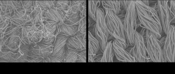
Scanning electron micrographs of 100% cotton twill on the left and 100% polyester knit on the right, captured by Hitachi S2600 SEM. Image courtesy of Sara Fleetwood, a PhD student at the BioProducts Institute in Dr. Johan Foster’s lab, Faculty of Applied Science. These images are part of Sara’s project on developing bio based hydrophobic […]
(Untitled)
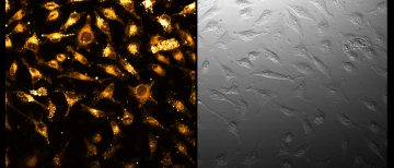
HeLa cells incubated with fluorescent organic nanoparticles for 24 hrs. Will Primrose, a PhD student in Dr. Zac Hudson lab, Dept of Chemistry, collected images using an Olympus FV1000 Laser Scanning Confocal microscope. Left image:Single photon images were captured with 405 nm laser light at 20% laser power with a 40x objective lens. Right image: […]
