(Untitled)
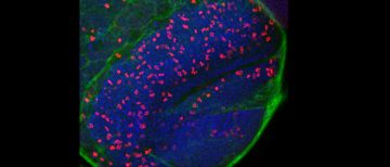
Image courtesy of Duo Cheng in Dr. Vanessa Auld lab, Dept of Zoology. Immunofluorescence labelling of cellular proliferation within the brain lobe of 3rd instar Drosophila melanogaster larvae. Green fluorescence represent glial cell membrane, red fluorescence represents the phosphohistone 3, marking cellular proliferation and blue is cell nuclei. The image was acquired by the Olympus […]
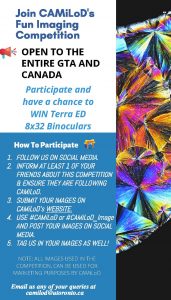
CAMiLoD’s Microscopy Imaging Competition Summer 2020
📢 Are you READY for CAMiLoD’S Summer Imaging Competition 2020? (It includes a SPECIAL GIFT for the Winner, Find Out Below) ➡️ PLEASE READ THE INSTRUCTIONS BELOW CAREFULLY TO ENSURE YOU FOLLOW THE RIGHT STEPS! 1: Go to https://www.camilod.ca/camilod-image-competition-2020 and fill all fields (Name, email, Institution and a brief description of your work of art) […]
(Untitled)
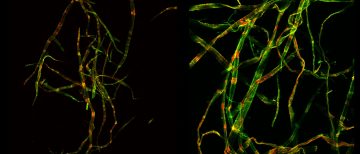
Immunofluorescence labelling of hardwood bleached kraft pulp fibers (left) and softwood bleached kraft pulp fibres (right). Image courtesy of Dr. Vinay Khatri (post-doctoral researcher) in Dr. Jack Saddler lab (Dept of Wood Science). The images were acquired with the Olympus FV1000 confocal microscope. Green fluorescence represents the crystalline cellulose and red fluorescence represents the amorphous […]
Congratulations to Miki, who won a Canadian Network of Scientific Platforms Scientist Award for 2020!
Congratulations to Miki for winning a Canadian Network of Scientific Platforms Scientist Award for 2020! Miki was given this award for her work as Research manager at BIF and the success of BIF as a scientific research platform. The BIF is open to everyone, and currently has 5 dedicated technical staff providing training and service. […]
BIF will re-open on an appointment-only basis on June 10th (Wednesday).
BIF will be operating on an appointment-only basis on June 10th (Wednesday). We ask all the BIF users to read BIF guidelines BIF guideline for re-opening (Phase 1) and to understand the additional precautions being taken during this time.
(Untitled)
Arabidopsis root that was fixed, cleared and stained with dyes. Mendel Perkins and Tommy Kuo in Dr. Lacey Samuels lab (Dept of Botany) use the Olympus BX53 fluorescence microscopy to perform image stitching and tiling.
BIF is closed until further notice.
BIF is closed as of March 18th 2020 until further notice.
(Untitled)
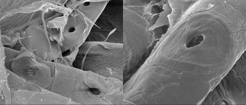
Scanning electron micrographs of softwood fibers showing irregular distribution and size of bordered pits. Image courtesy of Daniela Vargas Figueroa in Dr. James Olson lab (UBC Mechanical Engineering).
New Advances in Microscopy and Live Cell Imaging (ibidi Seminar) March 4th at 12pm LSI Rm 1330
Please join us for ibidi lunch and learn seminar on March 4th at 12pm in LSI Rm 1330.
Nikon MultiPhoton Imaging Talk on Feb 4th (Tuesday) at 1pm in BioSci Rm 4223
Nikon will give a lunch talk about Multiphoton Imaging Feb 4th (Tuesday) at 1-2pm in BioSci Rm 4223. Those interested in attending the demo can sign up at the talk and pizza lunch will be provided.