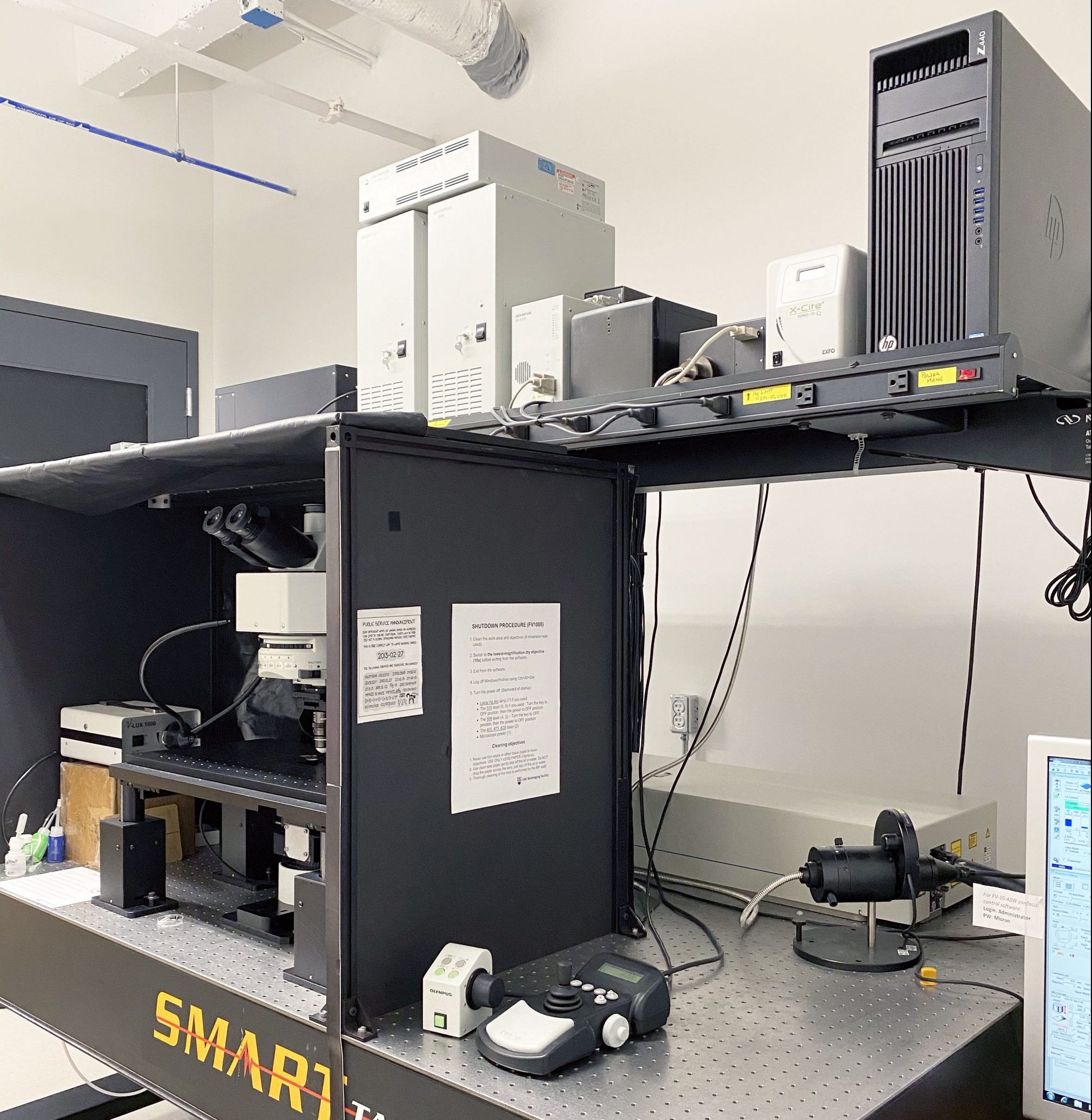
Capabilities:
– 3D multi-color confocal imaging/optical sectioning (4 channel detection)
– Two-photon imaging of thick samples (long working distance objectives and a fs pulsed, tunable laser, 690 – 1000 nm)
– Fluorescence Recovery After Photobleaching (FRAP)
– Real time data capture with simultaneous (dual scan units) image-scan for coincident bleach/photo-activation/photo-conversion while imaging
– Fluorescence spectra analysis
– Variable band width detectors for spectral analysis
Cost:
Regular confocal imaging: Academic users: $40/h, Industry users: $60/h
Two-photon imaging: Academic users: $66/h, Industry users: $150/h

Specifications:
Microscope:
Olympus BX61Wi upright microscope with epifluorescence illumination
Prior motorized stage (programmable sample repositioning or image tiling)
Conventional Confocal Imaging:
Olympus FV1000 confocal scan head with SIM (simultaneous) dual scan head for coincident imaging and photo stimulation/activation/bleaching
Solid-state laser lines for 405, 473, 514, 559, and 635 nm excitation
2 variable band-width tunable detectors, 2 detectors with conventional BP filters for 4 channel fluorescence detection
Forward propagation detector for ‘transmitted light’ image generation
High-Sensitive Detector (GaAsP) with 3 emission filter cubes (GFP/RFP, YFP/RFP, CFP/YFP)
Two-Photon Imaging:
Spectra-Physics MaiTai laser (690 – 1000 nm) with DeepSee dispersion compensation unit
Suitable for blue/green/yellow fluorescence protein imaging and second harmonic generation (SHG) imaging
3 or 4 NDD (non-descanned detector) channel back-propagated signal detection
2 NDD channel forward propagated signal detection
Filter cubes for NDD;
Blue, Green and Red – eg. DAPI, GFP and mCherry (Cube is marked MRVGR/VR)
Cyan, Yellow and Red – eg. CFP, YFP and mCherry (Cube is marked MRCYR/XR)
Conventional Objective Lenses:
10x/NA 0.4/Air
20x/NA 0.75/Air
40x/NA 0.95/Air
60x/NA 1.42/Oil
Objective Lenses for both conventional confocal and two-photon imaging:
30x/NA 1.05/Silicon oil (n=1.4); suitable for live cell imaging
40x/NA 1.25/Silicon oil (n=1.4); suitable for live-cell imaging
60x/NA 1.2/Water
Specialized Objective Lenses (Water Dipping Lenses):
10x/NA 0.6; WD 3mm; dedicated two-photon imaging
25x/NA 1.05; WD 2mm; dedicated two-photon imaging
40x/NA 0.8; WD 3.3mm; compatible for two-photon imaging
60x/NA 1.1; WD 1.5mm; compatible for two-photon imaging
Computer:
Olympus Fluoview software (FV10-ASW2) operated by Windows 7